Table of Contents
Alimentary Canal of Man or Digestive Tract:
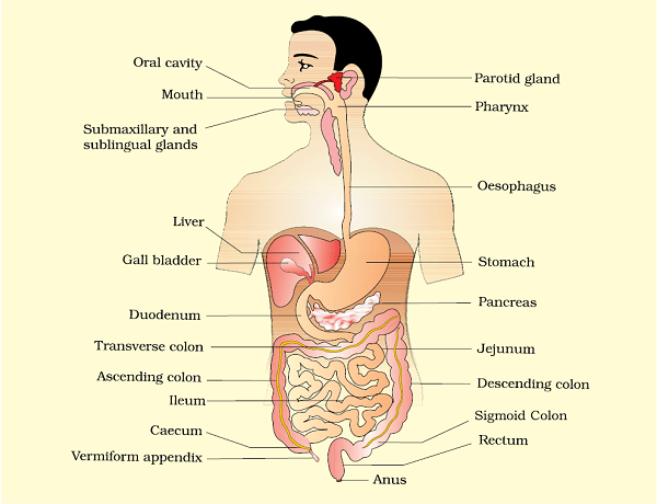
The human alimentary canal is a coiled and muscular tube about 9 meters long of varying diameters that possesses glandular epithelium. It opens anteriorly by means of mouth and terminates by another opening the anus. The various parts of the digestive tract or alimentary canal are the mouth, vestibule, oral or buccal cavity, oesophagus, stomach, small intestine, large intestine and anus.
Mouth:
The mouth is a transverse slit-like aperture which is bounded by two soft movable lips- upper lip and lower lip. The lips are covered with the skin on the outer side and lined with a mucous membrane on the inner side. The upper lip has a depression called philtrum, below the nose. The mouth is for ingestion and leads to the vestibule.
Vestibule:
It is a narrow space enclosed by lips cheeks externally and the gums and teeth internally. It is for temporary storage of food and leads to oral cavity.
Oral or Buccal Cavity:
It is a mouth cavity containing two lateral walls (cheeks), an anterior roof (hard palate), a posterior roof (soft palate) and a pair of jaws, the upper maxilla and lower mandible. The jaws contain the teeth. The tongue is a freely movable muscular organ attached to the floor of a buccal cavity by the frenulum and bears numerous taste buds in the taste papillae. Man bears 32 teeth that are heterodont and thecodont (embedded in the sockets of gums). These teeth occur in both the jaws. These teeth arise only twice in their lifetime. A set of temporary milk or deciduous teeth is replaced by a set of permanent or adult teeth. Man’s permanent teeth are heterodont and occur in four different types- incisors, canines, premolars and molars.
The oral cavity leads through the pharynx into the tube like oesophagus (food tube).
Functions of Tongue:
- It is an organ for receiving the sensation of taste.
- It is essential for phonation or speech along with lips, palate and teeth.
- It takes part in the ingestion of food.
- It brings the food below the teeth for mastication.
- Tongue wraps the food into a bolus and pushes it backwardly for swallowing.
- It takes part in cleaning of teeth.
Dental Formula:
The man possesses 32 permanent teeth and each jaw has 16 teeth each. In each half of jaws starting from middle and going backwards, there are two incisors, one canine, two premolars and three molars (2 + 1 + 2 + 3). Incisors are chisel-shaped and are used for cutting, chopping or gnawing. Canine is more pointed and used for ripping or shredding, premolars and molars are broad and used for shearing, crushing and grinding.
Dental formula of man: i 2/2, c 1/1, pm 2/2, m 3/3 = 32
Oesophagus:
It is a long (22-25 cm) narrow, muscular and tubular structure. It runs downward through the neck behind the trachea, passes through the diaphragm and opens in the stomach in the abdomen. Its inner mucosa is raised into longitudinal folds, oesophageal rugae, which close it to prevent the entry of air and also expand it to receive the food-bolus. It conducts the food to the stomach by peristalsis.
Oesophagus Functions:
- It conveys food from pharynx to stomach.
- Oesophagus allows the action of saliva to continue.
- It secretes mucus for lubrication.
- By remaining contracted during non passage of food, oesophagus prevents entry of air into the digestive tract.
Stomach:
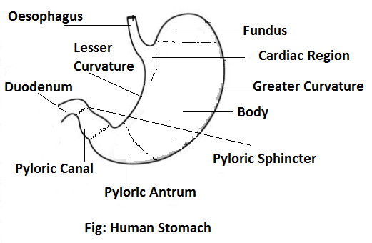
It is the most expanded part of the alimentary canal. It is a bag-like structure situated in the upper part of the abdomen towards the left side just under the diaphragm. It has two openings, two curvatures and three parts. The upper opening is called cardiac opening through which the stomach is connected to the oesophagus. The lower opening is called pyloric opening which has got pyloric sphincter. Towards the left side, the stomach has got greater curvature and towards the right side, it has lesser curvature. The fundus is the uppermost part of the stomach, the body is the central part of the stomach and pyloric portion is the lower part and it leads to the pyloric canal. The walls of the stomach have gastric glands which produce gastric juice which contains enzymes Rennin and Pepsin which help in digestion. The stomach also contains a small amount of HCl acid. It constitutes about 0.4% of gastric juice. The stomach also produces the castle’s intrinsic factor (a glycoprotein). This IF is essential for the absorption of Vitamin B12.
Function of HCl in the Stomach:
- It activates pepsinogen to pepsin and prorennin to rennin.
- It acts as an antiseptic and inhibits the growth of bacteria in the stomach.
- It inhibits the action of salivary amylase in the stomach.
- It provides an acidic medium in the stomach which enhances the action of gastric juice.
- It denatures many food proteins.
- It also controls the working of the pyloric sphincter.
Small Intestine:
It is the longest part of the alimentary canal which measures anything between 4.5 to 6 meters in length. It comprises three parts- duodenum, jejunum and ileum.
Duodenum:
It is the first part of the small intestine. It is somewhat C-shaped and about 25 cm long. The duodenum receives the hepatopancreatic ampulla, the opening of the common bile duct, a short tube formed by the union of the bile duct and the pancreatic duct.
Jejunum:
It is the middle coiled region of the small intestine which is 1.8-2.4 metres in length. Diameter is 4.0 cm. Wall is thicker, redder and more vascular. It is characterized by folds of membrane called Valvulae conniventes.
Ileum:
It is the longest part of the small intestine. It is about 7 m long and 2-3 cm in diameter that opens into caecum of the large intestine. The opening at the junction of ileum and caecum is guarded by a valve to prevent the regurgitation of food from the caecum. The ileum is highly coiled in the abdomen.
The inner surface of the small intestine is covered with numerous small villi which greatly increase the area for absorption. The wall of intestine contains numerous glands which secrete the intestinal juice containing several digestive enzymes. These villi are numerous in ileum of small intestine.
Peyer’s patches- Small white patches of lymphoid tissue, called Peyer’s patches, are found on the mucous membrane of the small intestine. They fight infection.
Functions of Small Intestine:
- Digestion of all digestible components of food is completed.
- Most of the absorption of digestive food occurs in the small intestine. For this, it has villi for increasing its absorptive surface by 10 times.
- It produces hormones like secretin, cholecystokinin, motilin, villikinin, enterogastrone, enterocrinin and duocrinin which control the secretory and absorptive activity of different parts.
Large Intestine:
It is much shorter than the small intestine and is basically for the absorption of water and for discharging the undigested wastes. It has three parts caecum, colon and rectum.
Caecum:
Caecum is a blind sac and is present where ileum joins the large intestine (just below the opening of ileum). In case of humans, it is very small and from it extends a small finger like vermiform appendix which gets infected during appendicitis. Caecum is large and spacious and a food storage organ in herbivores where cellulose digestion take place.
Colon:
It is the longest part of the large intestine which lies between caecum and rectum. Colon has three longitudinal bands of muscle fibres called taeniae coli. Contraction of these bands produces small pouches called haustra. Another characteristic of colon surface is the presence of small adipose projections called epiploic appendages. Colon has four parts-ascending, transverse, descending and sigmoid. Ascending colon is the first part present on the right side that moves upwards from the caecum. The transverse colon is the horizontal part placed transversely. Descending colon is the next region that moves down on the left side. The sigmoid colon is ‘S’ shaped and continues into the rectum. The sigmoid colon is also called pelvic colon because it occurs in the pelvic region. Food can remain in the colon for a long time, maybe as long as 36 hours before being passed out to rectum. Colon does not contain digestive glands. It harbours a number of micro-organisms which feed on the indigestible matter. Some of them are symbiotic. They produce K and B-complex vitamins. Villi and circular folds are absent.
Rectum:
It is a small muscular region at the end of the large intestine. It can store the undigested food for a very short time before passing it out through anus.
Anus:
It is the distal or terminal orifice of alimentary canal which lies in the inferior position between the buttocks. Anus is surrounded by two types of sphincters, internal and external. External sphincter is voluntary but closes the anus completely. Internal sphincter is involuntary. The closure is seldom perfect.


Comments (No)