Electron Microscopic Study of Cell and Organelles:
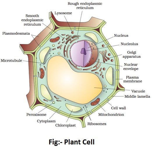
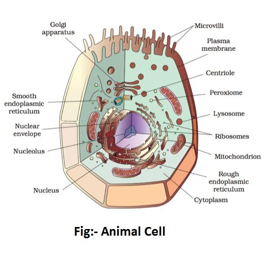
The invention of electron microscope and its subsequent use for scientific work has made it possible to have much clear and detailed picture of cell and organelles. For the purpose of electron microscopic study of cell and organelles, consider the electron micrograph shown by schematic diagram in the fig.


Under the electron microscope, the cell membrane is seen to be formed of three layers- a white layer, formed of liquid molecules, is sandwiched between two dark layers, formed of protein molecules. Three layers together measure approximately 75 Å in thickness. In plant cells, the cell membrane lies inside the cell wall.
The cytoplasm contains many structures. There is a complex and extensive network of membranes called the endoplasmic reticulum (ER). It is formed of paired membranes constituting a complex system of tubes, canals or channels. It is of two kinds- smooth and rough. The roughness of the membranes is because of the presence of some granules called ribosomes on their surface. Ribosomes are now regarded as the sites of protein synthesis.
Some of the membranes of endoplasmic reticulum open out in the cell membrane and the others in the nuclear membrane.
Golgi apparatus in cytoplasm appears, under an electron microscope, as a pile of 2-layered flattened sacs called cisternae or saccules, lying one within the other. The sacs have flattened or swollen ends near which lie the vesicles of varying size and shape. The sacs of Golgi apparatus appear like smooth endoplasmic reticulum and are often continuous with it at certain places.
Golgi apparatus stores the secretory substances and help in their transport.
Mitochondria are generally scattered throughout the cytoplasm. The electron microscope shows the mitochondria as vesicles bounded by two membranes and filled with a finely granular fluid called mitochondria matrix. The matrix has some ribosomes. There is a fluid material between the two membranes. The membranes of mitochondria resemble the cell membrane in structure and semipermeability. The mitochondria are formed of proteins, phospholipids, RNA and DNA. They are the sites of cellular respiration and regarded as the powerhouse of the cell.
There are some minute cell organelles inside the cell which resemble mitochondria in appearance. These are lysosomes. They store the digestive enzymes and prevent them from digesting the material of the cell itself. The lysosomes are referred to as suicide bags because on breaking up, they release the enzymes which digest and breakdown the cytoplasmic bodies and thus kill the cell.
Plastids are common in plants but absent in the majority of animal cells. They differ in shape, size, number, colour and arrangement in plant cells. They are of three types- chloroplasts (green coloured), leucoplasts (colourless) and chromoplasts (coloured other than green). They are formed of proteins, lipids and pigments.
Chloroplasts are responsible for photosynthesis in green plants, leucoplasts store food and chromoplasts give various colours to flowers, fruits and seeds.
The nucleus is the most important part of the cell. It plays an important role in the cell division and transfer of characters from parents to their offspring. It is surrounded by a definite wall termed the nuclear membrane. When seen under the electron microscope, the nuclear membrane is found to be double with a distant space between the inner and outer membranes. The important striking feature of the nuclear membrane is its ability to reappear after disappearance during the cell division.
- Mitosis: Process and Significance
- Meiosis: Process and Significance
- Chlorophyta- Green Algae
- Rhodophyta- Red Algae
- Bryophytes- Characteristics And Economic Importance
- Asexual Reproduction- Types, Characteristics And Significance
- Vegetative Propagation: Natural & Artificial Methods
- Mechanism or Working of Heart (Cardiac Cycle)
- Plant Kingdom-Tamil Board


Comments (No)