Table of Contents
Plant and Animal Cell: Important Points
- Cell was discovered by Robert Hooke in 1665.
- Cell is a structural and functional unit of living beings which consists of a membrane covered mass of protoplasm.
- Unicellular Organisms–
- Also called as acellular organisms.
- The body of the organism is made of a single cell.
- Example- Amoeba, Chlamydomonas, Paramecium, Chlorella, Acetabularia, Euglena.
- Amoeba does not have a fixed body shape and moves around by producing certain projections in the body called pseudopodia.
- Paramecium possesses tiny hair-like, structures distributed all over its body called cilia which help it in locomotion.
- Euglena and Chlamydomonas possess one or two whip-like locomotory structures called flagella.
- Multicellular Organisms–
- The body of the organisms is made of a many cells.
- Example- plants (metaphyta) and animals (metozoa).
- The cells are fewer and similar in coenobial or colonial organisms like Pandorina, Eudorina, Proterospongia and Volvox.
- Cells have diverse shapes. They may be flat, oval, spherical, cylindrical, cuboidal, spindle-shaped or irregular in shape.
- Guard cells found in leaves are bean shaped. Two guard cells are closely opposed to each other leaving a pore called stomata through which exchange of gases takes place.
- Cyanobacteria also called blue-green algae.
Plant and Animal Cell:
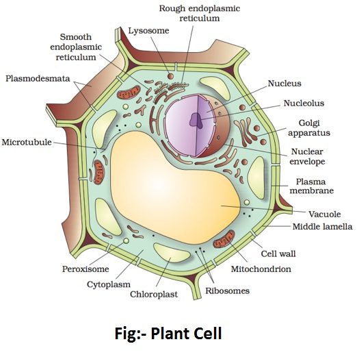
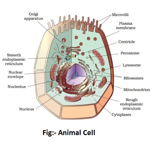
Cell wall:
- The cell wall is the outermost rigid protective covering found only in the plant cell.
- A plant cell wall is formed of three layers- middle lamella, primary cell wall, and secondary cell wall.
- The middle lamella is a thin, amorphous, intercellular cementing matrix present between adjacent cells. It is made up of pectates of calcium and magnesium.
- The primary cell wall is the layer present on the inner side of the middle lamella. It consists of a number of a loose network of cellulose microfibrils embedded in an amorphous gel-like matrix of hemicellulose and pectin. Cells such as parenchyma and meristems have only a primary wall.
- The secondary cell wall lies on the inner side of primary cell in the matured cells. It plays a key role in determining the shape of a cell. It is thick, inelastic, and is made up of cellulose and lignin.
Functions of Cell Wall:
- Cell wall provides shape to plant cells.
- It imparts rigidity to cells.
- It provides protection to the cell against any mechanical injury.
- Cell wall prevents the bursting of cells by maintaining osmotic pressure.
- It functions as an apoplast which is permeable to water and minerals dissolved in it.
- Cell growth is made possible only when the cell wall undergoes extension.
- It provides strength to the aerial part of the plant body to withstand gravitational forces.
- It sometimes performs the function of offense and defense.
Cell membrane:
- The cell membrane is a delicate covering that surrounds the jelly-like cytoplasm present inside the cell.
- In animal cells, it is the outermost layer while in plant cells it is surrounded by the cell wall on the outside.
- It is a living structure and has tiny holes which allow materials to move in and out of the cell.
- It is made up of lipids of proteins.
- The cell membrane is also called plasma membrane.
Cytoplasm:
- Cytoplasm is a complex colloidal, viscous, semi-transparent, jelly-like substance.
- It contains several structures called cell organelles floating in it except the nucleus.
- It is limited by a plasma membrane or cell membrane.
- It also contains nucleotides, vitamins, tRna, enzymes, sugar and amino acids, etc.
Functions of Cytoplasm:
- It provides raw material to the cell organelles for their functioning.
- It helps movement of the cellular materials around the cell through a process called cytoplasmic streaming.
- It serves as the seat for the synthesis of fats, nucleotides, some carbohydrates and proteins etc.
- It performs various catabolic activities like glycolysis and anaerobic respiration etc.
Cell Organelles:
- Cell organelles are extremely small structures present in the cytoplasm and concerned with cell function.
- Following cell organelles are commonly found in cells.
Endoplasmic Reticulum:
- The largest of the internal membranes is called the endoplasmic reticulum (ER).
- The name endoplasmic reticulum was given by K.R.Porter.
- Endoplasmic Reticulum is a network of tubular channels present throughout the cytoplasm.
- It is found in the liver, pancreas, brain, adipose cells, and in spermatocytes.
- When ribosomes are present in the outer surface of the membrane it is called as rough endoplasmic reticulum (RER). It occurs in almost all the cells and is actively busy in the synthesis of proteins and enzymes. It occupies the central position and so remains connected with the nuclear envelope.
- When the ribosomes are absent in the endoplasmic reticulum it is called as smooth Endoplasmic reticulum (SER). It is mainly engaged in the synthesis of steroids, glycogen, and lipids. It occupies the peripheral position and so remains continued with the plasma membrane.
Endoplasmic Reticulum is formed of three types of elements-
- Cisternae– are elongated, flattened, two-layered, and unbranched tubular elements arranged in a parallel row. They are mainly found in the protein forming cells of the liver, pancreas, and brain, etc. They are basophilic in nature.
- Vesicles– are the rounded and spherical elements which are found scattered in the cytoplasm. These are studded with ribosomes and can be seen in liver and pancreatic cells.
- Tubules– are irregular in shape, branched, smooth-walled, enclose a space. These occur in cells which are busy in the synthesis of steroids.
Functions of Endoplasmic Reticulum:
- Endoplasmic Reticulum acts as a cell circulatory system and transports the material through the cytoplasm because it remains connected with the nuclear membrane on one side and the cell membrane on the other side.
- It provides supplementary mechanical support to the colloidal matrix of cytoplasm as well as gives support and shape to the cell.
- It increases the surface area for cellular activities.
- It helps in the formation of primary lysosomes and nuclear envelope during cell division.
- Endoplasmic Reticulum provides membranes to the Golgi apparatus for the production of vesicles and Golgi vacuoles.
- Enzymes located over the smooth Endoplasmic reticulum of liver cells take part in the breakdown of glycogen.
- The smooth Endoplasmic reticulum takes part in fat synthesis inside adipose cells.
- The smooth Endoplasmic reticulum takes part in the synthesis of steroid hormones. Example- testosterone in testis, estrogens in the ovary.
- In skeletal muscles, smooth Endoplasmic reticulum takes part in excitation contraction coupling.
- The rough endoplasmic reticulum are the site for protein synthesis.
- The rough endoplasmic reticulum forms smooth Endoplasmic reticulum by avoiding ribosomes.
Golgi Apparatus or Golgi Complex:
- Camillo Golgi, an Italian scientist discovered the Golgi apparatus in 1898.
- Golgi apparatus is a stack of flat membrane enclosed sacs. It consists of cisternae, tubules, vesicles, and Golgi vacuoles, and it helps in secretion, membrane transformation, and synthesis of complex biochemicals.
- It is found in all eukaryotic cells with the exception of mammalian erythrocytes, sieve tube elements, sperms of bryophytes, and pteridophytes. Prokaryotic cells do not contain the apparatus.
- In plant cells and invertebrates, the Golgi Apparatus exists in the form of unconnected units, called dictyosomes. These unconnected stacks of flattened sacs (cisternae) are found to be scattered irregularly in the cytoplasm. These cisternae vary in their number and each of them is really a stack of Golgi complex. The dictyosomes help to synthesize pectin and carbohydrates in the plant cells.
Functions of Golgi Apparatus:
- It helps in the formation of secretory vesicles. The cellular secretions like proteins are concentrated into membrane bound zymogen granules which arise from the maturing faces of Golgi cisternae.
- It helps in the formation of cell wall by the synthesis of polysaccharides like pectin, hemicellulose during cytokinesis.
- It is also involved in storage, condensation, packaging, and transfer of materials.
- The Acrosome is a special structure present near the tip of sperm that contains hydrolytic enzymes for digesting the protective coverings of the egg. The acrosome is formed by the Golgi complex. After the formation of the acrosome, the Golgi apparatus degenerates.
- It synthesizes glycoprotein by combining proteins from rough endoplasmic reticulum and carbohydrate.
- It synthesizes various complex polysaccharides like mucopolysaccharides, hemicellulose, and cellulose, etc.
- The secretory vesicles or vacuoles of Golgi complex function as primary lysosome by storing digestive enzymes in the inactive state.
- It also secretes mucilage and gums etc.
- It helps in the vitellogenesis and in the origin of pigments like melanin.
- Hormones synthesis of endocrine glands is mediated through Golgi complex.
Mitochondria:
- Mitochondria are tiny rod-like structures found in the cytoplasm.
- Also known as the power house of cell, because during the oxidation of food, the energy released is stored in the ATP (Adenosine triphosphate) molecules.
- They were named as bioblast by Altman (1894) but Benda (1897) assigned the present name mitochondria.
- A mitochondrion is generally cylindrical in outline and is surrounded by the membranes- outer and inner membranes.
- The outer membrane is smooth, highly permeable to small molecules and it contains proteins called Porins.
- The inner membrane is selectively permeable and remains rich in enzymes and carrier proteins. It projects into the mitochondrial cavity in the form of a finger-like projection called cristae. These cristae increase the surface area for cellular respiration.
- The outer and inner membranes are separated by a space of 60-80 A° and are called an outer compartment or perimitochondrial space.
- The inner mitochondrial membrane encloses the inner compartment which remains filled with proteinaceous material called mitochondrial matrix. It remains rich in proteins, lipids, circular DNA, 70s ribosomes, and certain granules.
- The elementary particles or oxisomes, the stalk particles like tennis racket (F0−F1) combination are found on the inner membrane of the mitochondrion.
Functions of Mitochondria:
- Mitochondria perform the Krebs cycle or tricarboxylic acid cycle of aerobic respiration. Oxygen is used as a terminal oxidant. CO2 and H2O are produced.
- Mitochondria carries enzymes for the electron transport system as well as for oxidative phosphorylation.
- A number of fatty acids are synthesized in the matrix of mitochondria. Mitochondria also possess enzymes for their elongation.
- Mitochondria contain DNA and hence help in extranuclear or cytoplasmic inheritance.
Ribosomes:
- Ribosomes are the nucleoprotein protoplasmic structures which are found both in prokaryotic and eukaryotic cells except in red blood cells of mammals. They are found scattered freely in the cytoplasm and attached to Endoplasmic Reticulum. They also occur in mitochondria and plastids.
- The ribosome consists of RNA and protein: RNA 60 % and protein 40%. During protein synthesis, aggregation of ribosomes occurs which attach to the same mRNA and forms the polysomes or polyribosomes.
- The function of polysomes is the formation of several copies of a particular polypeptide during protein synthesis.
- They are free in non-protein synthesizing cells.
- In protein synthesizing cells they are linked together with the help of Mg2+ ions.
Lysosomes:
- Lysosomes were discovered by Christian de Duve (1953).
- Lysosomes are tiny sacs containing digestive enzymes and are often called suicide bags because under the adverse conditions they can eat the very cell in which they are present.
- Lysosomes are formed directly from the Endoplasmic reticulum or Golgi complex.
Functions of Lysosome:
- They digest the large extracellular molecules of food (phagosomes or pinosomes) which enter the cell. The lysosomes of WBCs help the later to engulf foreign particles.
- During the favourable starvation, the lysosomes digest the food material like protein, liquids, and glycogen stored within the cells (autophagy, intracellular scavenging) and provide energy to the cell.
- Lysosomes can digest the entire cell in which they are present (autolysis). This takes place when the cell is about to die.
- Lysosomes bring about the extracellular digestion by secreting hydrolytic enzymes outside the cells. The lysosomes of human sperms secrete enzymes (protease and hyaluronidase) outside the cell to dissolve the barrier membranes of an ovum and to facilitate the process of fertilization.
- They help in differentiation and metamorphosis– degeneration of the tail, fins, and gills of tadpole and later absorbed by the other cells.
- They bring about the hydrolysis of proteins and starch during seed germination.
Centrosome:
- Centrosome is a small body present close to the nucleus in animal cells.
- It contains two tiny granular bodies called centrioles.
- The centrioles help in cell division.
Plastids:
- Plastids are the special cytoplasmic organelles found in eukaryotic plant cells and in some protozoans like Euglena. They perform the function of synthesis of food and storage of carbohydrates, lipids and proteins.
Depending on their colour, the plastids can be categorised as under:
Chloroplasts-
- Chloroplasts are green plastids. It is an important plastid because they contain a green pigment called chlorophyll which manufactures food for the plant using solar energy through the process of photosynthesis. Produce starch and found in leaves.
- It has the protein synthetic mechanism as DNA of chloroplast codes for chloroplast rRNA, tRNA, and ribosomal proteins.
- They are called as food factory of cell because it synthesizes the ATP and NADH2 by cyclic and non-cyclic photophosphorylation. It performs the anabolic reaction and synthesizes the food.
- Chloroplasts show phototactic movements. In some algae, they possess stigma or eye spot for providing photosensitivity to motile structures.
Leucoplasts-
- These are Colourless plastids and help in the storage of food in various forms. They may be amyloplasts (starch storing), elaioplasts (lipid storing), and proteinoplasts or aleuroplasts (protein storing). Leucoplasts are found in the cells of storage tissue, endosperm, embryo, and germ cells.
Chromoplasts–
- Yellow, orange, or red-coloured plastids containing various pigments like carotenes and xanthophyll, etc. They impart colours to the flowers and ripe fruits. For example- carotene gives the red colour to the tomato fruit.
Vacuole:
- A space within the cytoplasm of a plant cell, containing cell sap.
- Animal cells normally have many small vacuoles while plant cells generally have one big vacuole.
- Provide turgidity and rigidity to plant cells.
Nucleus:
- The nucleus is a dense body found generally in the center of the cell.
- It is the largest among all cell organelles.
- It may be spherical, cuboidal, ellipsoidal, or discoidal.
- It has a double-layered covering- the nuclear membrane which encloses a jelly-like ground substance.
Function of Nucleus:
- It is essential for the survival and long term continuation of cells.
- It maintains all the metabolic activities of the cell, cell maintenance, and cell replication.
- It is involved in maintaining regular cell division and differentiation.
- It regulates the inheritance of characters from generation to generation.
- Coding the information from DNA for the production of enzymes and proteins.
- DNA duplication and transcription takes place in the nucleus.
- In nucleolus ribosomal biogenesis takes place.
- Mitosis: Process and Significance
- Meiosis: Process and Significance
- Alimentary Canal of Man or Digestive Tract
- Mechanism or Working of Heart (Cardiac Cycle)
- Asexual Reproduction- Types, Characteristics And Significance
- Vegetative Propagation: Natural & Artificial Methods
- Male Reproductive System
- Female Reproductive System
- Gonads (Ovaries in Females and Testes in Male)
- Thyroid Gland: Important Hormones And Deficiency Disorder
- Theories of Evolution: From Tamil Board Book




Comments (No)