Table of Contents
Respiratory organs present in human being:

Nasal Cavity:
Nasal Cavity is a hollow structure partitioned into two nasal chambers by the nasal septum. It is composed of cartilage, bone, connective tissue and muscles. There are pairs of openings situated at the anterior and posterior ends of the nasal chambers. The anterior nostrils lead to the nasal cavity.
Pharynx:
It is a vertical tube which extends between the cranial base (above the level of the soft palate) and middle of the neck. It has three parts- nasopharynx, oropharynx and laryngopharynx. The nasopharynx is the upper part that lies above the level of the soft palate. Internal nares open into it. There are two other openings of eustachian tubes. The oropharynx is the middle part which is a continuation of the buccal cavity. Laryngopharynx or hypopharynx is the lower part. It is a common passage for food and air. Gullet or opening of the oesophagus is towards the backside while glottis or opening of the larynx is in front.
Larynx:
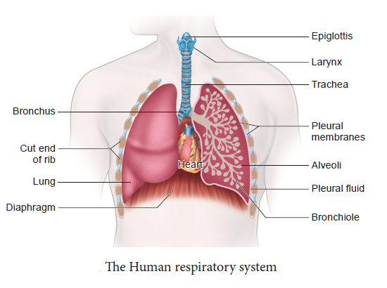
It is also called the voice box or Adam’s apple. It is situated at the anterior part of the trachea and communicates with the orophyarnx by a slit-like aperture, the glottis. Stiff leaf-like cartilage, the epiglottis, protects the glottis during swallowing. The larynx is roughly tubular, elongated structure and it is composed of nine cartilages, connective tissue, extrinsic and intrinsic muscles lined with mucous membrane. The larynx proper contains pairs of shelf-like folds- superior folds (fake vocal cords) and ventricular (true vocal cords). The space between true vocal cords is known as glottis. The vocal cords set into motion as expired air is forced between them through the glottal opening and sound is produced.
Trachea or Wind Pipe:
The trachea is a thin-walled, long tubular structure which runs downward through the neck in front of the oesophagus. It is about 11 cm long and 2.5 cm wide. It is supported by 16-20 cartilaginous rings called tracheal rings. These are C-shaped, posteriorly incomplete rings which prevent the trachea from collapsing during inspiration and provide strength and flexibility.
Histologically, trachea comprises an internal ciliated columnar epithelium bearing glandular cells (mucous cells) connective tissue containing cartilages, blood vessels and nerve fibres. The mucus keeps the walls of trachea moist and traps the dust particles.
Bronchi:
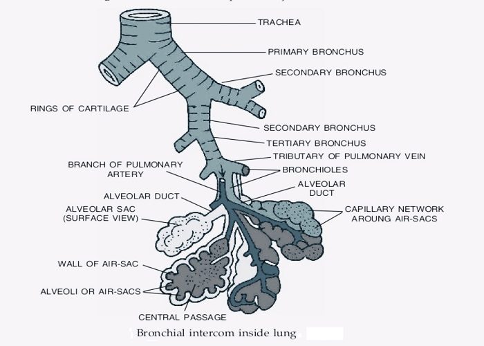
Inside the thorax, the trachea bifurcates into two bronchi at the level of 5th thoracic vertebra and each of which enters into one lung. In each lung, the bronchus again redivides into numerous small branches called bronchioles. These bronchioles are without cartilaginous rings and again redivide into the finer terminal bronchioles within each lobule. The terminal bronchioles at their ends continue to respiratory bronchioles, which are short, tubular and lined with ciliated columnar epithelium without goblet cells. A short distance along their course, they again branch and give rise to 2-11 radiating alveolar ducts.
Each alveolar duct is a long, thin-walled tubular structure which gives off a number of branches in the form of pouches as alveolar sacs. The alveolar sacs open into alveoli. The alveoli are extremely thin-walled, polyhedral sacs richly supplied with blood capillaries. There are about 300 millions of alveoli in the two lungs. The blood is separated from the alveolar air by two thin layers of cells- the alveolar epithelium and the capillary endothelium and the gaseous exchange occurs through these two layers.

Lungs:
The lungs are housed on either side of the heart in the pleural cavities of the thoracic cavity and are connected to the pharynx by trachea. The lungs are covered by extremely thin double membrane sacs called pleural sacs. The outer membrane of the pleural sac is called parietal pleura (in close contact with thoracic lining) while the inner membrane is known as visceral or pulmonary pleura (in contact with lung surface). Between these layers, there is a narrow pleural cavity filled with a watery pleural fluid which performs two functions- firstly, allows free frictionless movements of lungs and secondly, protects the lungs from mechanical shocks. The mediastinum is the partition between the two lungs. It includes the pleura of both sides. It is generally defined as the interval between two pleural sacs. In the lower region, the two pleura separate to leave a cavity, the mediastinal space, for the heart.
Lungs are soft, spongy, elastic and subconical sacs. Each lung has a triangular depression called hilus or hilum. Here, various structures enter or leave the lung. They are pinkish in colour at birth but gradually turn greyish with age under the influence of environmental pollutants. The lungs are divided externally into lobes by fissures. The left lung has two lobes, left superior and left inferior demarcated by an oblique fissure. The right lung has three lobes, right superior, right middle and right inferior demarcated by horizontal and oblique fissures. The left lung is smaller than the right lung. On average, an adult right lung weighs 625 gm, while the left lung weighs 565 gms.
The lungs are situated in the thoracic chamber which is anatomically an air-tight chamber. The thoracic chamber is formed dorsally by the vertebral column, ventrally by the sternum, laterally by the ribs and on the lower side by the dome-shaped diaphragm. The anatomical set up of lungs in the thorax is such that any change in the volume of the thoracic cavity will be reflected in the lungs (pulmonary) cavity. Such an arrangement is essential for breathing as one can not directly change the pulmonary volume.


Comments (No)