Table of Contents
Mechanism of Stomatal Movement:
What is Stomata?
Stomata help in gaseous exchange at the time of respiration and photosynthesis. They are very minute apertures, usually found on the epidermis of the leaves. Each stoma is surrounded by two kidney-shaped special epidermal cells, known as guard cells. The wall of guard cells near the pore is thick. The outer wall is thin, elastic and semipermeable.
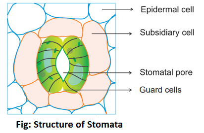
The stomata may be found in all the aerial parts of the plant. They are never found on its roots. Usually, the stomata are found scattered on the dicotyledonous leaves whereas they are arranged in parallel rows in the case of monocotyledonous leaves.
The stomata may be found on both the surfaces of the leaf, but their number is always greater on the lower surface. However, the upper surface of the leaves of banyan and rubber trees lack stomata. The upper surface of the leaves of several xerophytes also lacks stomata. The free-floating leaves of the water plants bear stomata only on their upper surface.
In normal condition, the stomata remain closed in the absence of light. They are always open in the day time or in the presence of light.
Mechanism of Stomatal Movement (Opening and Closing of Stomata):
A variety of external and internal stimuli affect the opening and closing of stomata by altering the size of stomatal pores. The important among them are light and dark, CO2 concentration, water supply, pH of the cell sap etc. In most of the cases when the water supply is adequate, the stomata tend to open during the day time in response to light and close at night. The opening and closing of stomata operate as a result of turgor changes in the guard cells. When the guard cells become turgid, their thin walls get extended and thick walls become slightly concave so that the stomatal aperture opens. On the other hand, the guard cells become flaccid when they lose water. Their thick walls revert back to their original position resulting in the closure of the stomatal pore.
Three theories regarding the opening and closing of stomata are as follows:
Guard Cells Photosynthesis:
Schwendener (1881) thinks that chloroplasts of guard cells perform photosynthesis in light and produce sugar. The raised sugar level increases the osmotic concentration of guard cells. Guard cells absorb water, swell up and develop a pore in between them. In the darkness, photosynthesis stops and the sugar is transported out of the guard cells.
Starch Sugar Hypothesis:
This theory was formulated in 1923 by J.D. Sayre and modified by Steward in 1964. An outline of this theory is given below-
In Light:
- Photosynthesis occurs in the light which consumes the respiratory CO2 present in the intercellular spaces.
- This lowers the H+ concentration of cell sap and the pH of guard cells is increased.
- High pH favours the activity of enzyme phosphorylase which converts starch into glucose-1-phosphate-
| Starch ——–Phosphorylase————–pH 7.0——–> Glucose-1-phosphate |
- The glucose-1-phosphate is further converted to glucose-6-phosphate by enzyme phosphoglucomutase-
| Glucose-1-phosphate ⇌ Glucose-6-phosphate |
- The glucose-6-phosphate is converted to glucose and phosphate by enzyme phosphatase-
| Glucose-6-phosphate ——————> Glucose + Phosphate |
- The glucose and phosphate units dissolve in the medium and increase the concentration of cell sap-
- This cause further increases in the osmotic pressure of guard cells and consequently, its DPD (Diffusion pressure deficit) is also increased. This results in the movement of water into the guard cells from surrounding cells. The guard cells become turgid and swell. Thus, the stomatal pore becomes open.
In Dark:
- Photosynthesis does not occur in dark. The level of respiratory CO2 in the substomatal cavity is increased. This results decrease in the pH of guard cells.
- At low pH, the glucose-molecules are converted to glucose-1-phosphate making use of respiratory ATP in presence of enzyme hexokinase-
| Glucose + ATP ——Hexokinase——————> Glucose-1-phosphate + ADP |
- The glucose-1-phosphate molecules are converted to starch in presence of enzyme phosphorylase-
| Glucose-1-phosphate —————> Starch |
- Synthesis of starch leads to the dilution of cell sap by consuming its dissolved glucose molecules. Thus the osmotic pressure of cell sap is decreased and its DPD is also decreased. The guard cells then lose water to the surrounding cells and become flaccid. The stomatal pore is then closed.
K+ Ion Pump Hypothesis:
This theory was proposed by Levitt (1974) and elaborated by Raschke (1975) and Bowling (1976). A brief account of this theory is given below-
In Light:
- The starch content of the guard cells disappears in the light. First of all the starch is converted into organic acids particularly phosphoenol pyruvic acid. Phosphoenol pyruvic acid then combines with CO2 to produce oxalo acetic acid and then malic acid.
- The organic acids, viz., malic acid, dissociate into malate anion and H+ in the guard cells.
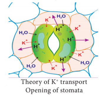
- H+ is transported to epidermal cells particularly the subsidiary cells (if present) and K+ are taken into the guard cells in exchange of H+. The process is called ion exchange.
- K+ are balanced by organic anions (i.e. malate). Some Cl– ions are also taken in to neutralize a small percentage of K+.
- H+ – K+ exchange is an active process that requires the involvement of energy (ATP) supplied either by respiration or photophosphorylation.
- Increased concentration of K+ and malate ions in the vacuole of guard cells causes sufficient osmotic pressure to absorb water from surrounding cells.
- Increased turgor of guard cells due to entry of water results stomatal pore to open.
In Dark:
- Carbon dioxide concentration is increased in dark in the sub-stomatal cavity because photosynthesis is stopped but respiration is continued.
- A higher concentration of CO2 in the substomatal cavity prevents proton gradient across the protoplasmic membrane in guard cells. As a result, the active transport of K+ into guard cells stops.
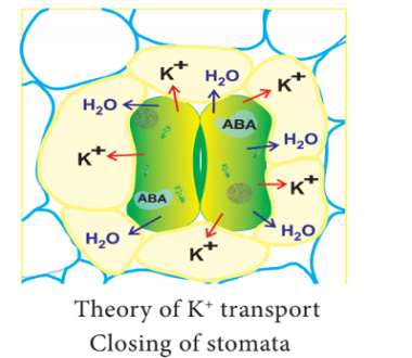
- Cowan et al. (1982) proposed that the closure mechanism involves the participation of an inhibitor hormone abscisic acid, which functions at lower pH. As soon as the pH of guard cells decreases, the abscisic acid inhibits K+ uptake by changing the diffusion and permeability of the guard cells.
- Malate ions present in the guard cell cytoplasm combine with H+ to produce malic acid. Excess of malic acid inhibits its own synthesis by decreasing the activity of PEP-Carboxylase.
- These changes cause reversal of ion movement so that K+ is transported out of guard cells into surrounding epidermal cells.
- The osmotic pressure of guard cells is thus decreased.
- This results movement of water from guard cells to surrounding cells.
- The guard cells become flaccid and the stomatal pore gets closed.




Comments (No)