Spinal Cord:

Location:
The spinal cord is a cylindrical cord-like structure that lies in the neural canal of the vertebral column. It extends downwards from the brain stem to the lumbar region. Like a brain, it is also protected by three meninges, cerebrospinal fluid and a cushion of adipose tissue.
Morphology:
The spinal cord is cylindrical with two swellings: one in the cervical region and the other in the lumbar region. It extends from the lower end of the medulla oblongata to the first lumbar vertebra where it first tapers to a point, called conus medullaris, but then becomes non-nervous and thread-like, called filum terminale present in the coccyx.
Structure:
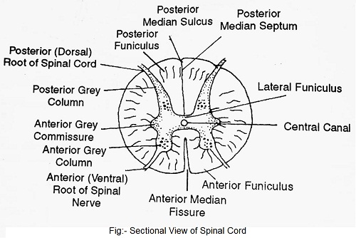
The central canal of the spinal cord is a continuation of the fourth ventricle. It is filled with cerebrospinal fluid. Two deep median grooves namely, dorsal fissure and ventral fissure divide the spinal cord into two symmetrical halves. Unlike the brain, in the spinal cord, grey matter forms the inner core. It appears H-shaped in cross-section and contains cell bodies of association neurons or interneurons and motor neurons. In a cross-section of the spinal cord, its grey matter appears penetrating into the white matter and forms horns on either lateral side. These are called dorsal horns, ventral bones and lateral horns.
The white matter surrounds the grey matter. In each half of the spinal cord, the white matter is divided into four columns or funiculi. These are one anterior, one posterior and two lateral funiculi. Each funiculus is formed of nerve tracts. A nerve tract is a bundle of nerve fibres originating and terminating at similar sites. These contain ascending as well as descending tracts.
(1) Ascending nerve tracts conduct nerve impulses from the spinal cord to the brain. The sensory impulses from the peripheral sense organs are brought to the spinal cord by sensory spinal nerves and from here through these tracts are conveyed to the brain.
(2) Descending nerve tracts conduct impulses from the brain to different levels of the spinal cord. The motor impulses from the brain are transmitted to the cells of the ventral and lateral horns of the spinal cord. The axons of these cells conduct the motor impulses through spinal nerves to effector organs (glands and muscles).
Functions- Spinal cord performs sensory, motor and reflex functions.
(1) It is the centre for many reflexes (spinal reflexes). Effectors present on the skin surface receive the external stimuli which are received by posterior root ganglion through sensory or afferent nerves. It is converted to motor impulse by convertor or interneurons and sent to effector on the skin through motor or efferent nerves. Thus, the spinal cord aids the brain in its functions.
(2) It provides nerve connection to a large number of body parts.
(3) It conducts sensory and motor impulses to and from the brain.


Comments (No)