Table of Contents
Female Reproductive System:
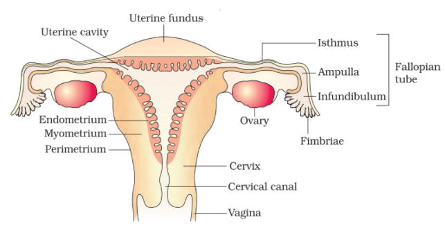
The sex organs of female mammals are specialized for egg production, for receiving sperms from the male, for providing an adequate medium for fertilization and for protecting and nourishing the developing embryo. For these multifarious activities, the reproductive system of human female consists of two ovaries, Fallopian tubes (oviducts), uterus, vagina and external genitalia.
Ovaries:
There are two ovaries situated in the abdominal cavity one on either side of the vertebral column. Each ovary is roughly almond-shaped measuring 3 cm in length, 1.5 cm in breadth and 1 cm in thickness. Each ovary is attached to the broad ligament of the abdominal wall by the mesovarium and to the uterus by a double fold of peritoneum, called an ovarian ligament. Both the ovaries lie in the abdomen and they produce ova or eggs by the process of oogenesis and also secrete two important female sex hormones called Oestrogen and Progesterone. Generally, alternate ovaries liberate only one ovum in each menstrual cycle (average duration, 28 days). A human female produces only about 450 ova over the entire span of her reproductive life which lasts about 40-50 years. Although follicular cells and oocytes undergo degeneration during follicular atresia, some theca cells formed from the stroma and located around the follicular, persist and become active. These are called interstitial cells. They secrete a small amount of androgen.

Fallopian Tubes/ Oviducts/ Uterine Tubes:
They are a pair of muscular and internally ciliated tubes of 10-12 cm length which arise near the ovary and ends at the uterus. A fallopian tube is differentiated into four parts-
- Infundibulum- Funnel-shaped free end, fimbriated and lies near the ovary. It bears a pore or ostium for entry of ovum.
- Ampulla- A slightly swollen and curved part behind infundibulum where fertilization of ovum takes place.
- Isthmus- It is the narrow and straight part of the fallopian tube.
- Uterine Part- It is about 1 cm long part that passes into the uterine wall. Passage of ovum is facilitated by movements of cilia and muscular contractions of the wall.
In most vertebrates both the ovaries and oviducts are functional. In birds, the right ovary and right oviduct are atrophied. Being non-mammalian, the birds also lack mammalian sex organs and characters like uterus, external genitalia and mammary glands.
Uterus:
It is a large, pear-shaped, muscular and thick-walled organ, measuring about 8 cm long, 5 cm broad and 2 cm thick and connected on its either sides with the Fallopian tubes. The uterus consists of an upper saccular portion, the body and a lower constricted portion, called the cervix. The body of uterus comprises three coats: the innermost endometrium, middle myometrium and outermost perimetrium. The endometrium is glandular containing specially designed blood vessels and uterine glands. The myometrium is muscular having layers of smooth muscle fibres. The perimetrium has a serous membrane and thin connective tissue. Cervix is mainly a sphincter muscle that closes the uterine lumen and prevents the entry of foreign particles into the uterus. Various important functions of the uterus, i.e., menstruation, pregnancy and labour, are performed by its different layers.
Vagina:
It is a tubular female copulatory organ, a passageway for menstrual flow as well as the birth canal of about 8 cm length between external opening (vaginal orifice) in vestibule and cervix with depression or fornix around cervix and transverse folds or vaginal rugae. Its mucous membrane is non keratinized stratified squamous epithelium with glands at places. In virgins, the vaginal orifice is partially covered by an annular centrally perforate membrane called the hymen.
External Genitalia/ Vulva:
The external genitalia of human female comprises mons veneris (the fatty mounds or pubis), labia majora, labia minora, clitoris, urinary meatus, vaginal orifice and Bartholin’s glands. On the pubis region, coarse hairs appear at puberty and persist throughout life. The labia majora (large lips) are paired rounded fleshy folds of skin which extend down from the pubis and surround the vaginal opening. The labia majora corresponds to the scrotum of the male and contains hair externally and sweat and sebaceous glands internally. The labia minora (small lips) are two small folds of mucous membrane situated on the inner side of labia majora. They are devoid of hair and glands. At the upper end, the folds meet to form a thin covering, the prepuce. The clitoris is a small erectile organ which is highly sensitive and is located at the junction of the labia minora, below the prepuce. The clitoris corresponds developmentally to the penis of the male. The urinary meatus is a small opening of the urethra below the clitoris and above the vaginal orifice that is the external opening of the vagina through the hymen. There are two bean-shaped glands on either side of the vaginal orifice. These glands are comparable to the bulbourethral glands of the male. Each of them opens into the space between the hymen and the labia minora through a duct and secretes a lubricating fluid.


Comments (No)